Comprehensive Radiology Services
Northern Nevada Medical Center offers comprehensive radiology services to residents of Sparks, Reno and the surrounding region. Among the services offered are:
- Diagnostic radiology
- Fluoroscopy
- Computed tomography (CT)
- Computed tomography angiography (CTA)
- Magnetic resonance imaging (MRI)
- Diagnostic ultrasound
- Vascular ultrasound
- Nuclear medicine
- Mammography
- Bone densitometry (DEXA)
- Cardiovascular imaging lab
- Interventional radiology (angiography)
- Heart catheterization
- Percutaneous cardiac intervention (PCI)
- Electrophysiological study (EPS)
For Diagnostic Imaging scheduling, call 775-356-4042.
 Nationally Accredited in Radiology Services
Northern Nevada Medical Center is accredited in mammography, CT, MRI and ultrasound/vascular imaging services by the American College of Radiology.
Prepare for Your Imaging Test
Learn more about what to expect before, during and after your imaging procedure.
Advanced Imaging Technology
The advanced diagnostic imaging technology at Northern Nevada Medical Center gets results to your physician quickly, so he or she can make a diagnosis and recommend treatment for you as soon as possible. Your physician can access your results immediately, from any location, through our secure imaging web site. Imaging technology at Northern Nevada Medical Center includes:
X-ray
X-ray images are produced when a small amount of radiation passes through the body to create an image on sensitive digital plates on the other side of the body. The ability of X-rays to penetrate tissues and bones depends on the tissue's composition and mass, and the difference between these two elements creates the image. Contrast agents, such as barium, may be swallowed to outline the esophagus, stomach and intestines to help provide better images of an organ.
Computed Tomography (CT)
Computed tomography, also known as a CT or CAT scan, generates detailed images of an organ by using an X-ray beam to take images of many thin slices of that organ and joining them together to produce a single image. The source of the X-ray beam circles around the patient and the X-rays that pass through the body are detected by an array of sensors. Information from the sensors is computer processed and displayed as an image on a high resolution monitor.
Computed Tomography Angiography (CTA)
A CT scanner can be used to obtain images of blood vessels noninvasively. Iodine is a contrast material that may be injected into a vein using a small intravenous needle, creating no need for invasive catheter placement. This procedure can be used to screen for arterial diseases such as aortic dissection, carotid stenosis, aneurysms, stroke and vascular disease of the kidney. Resulting images may display the anatomical detail of blood vessels more precisely than an ultrasound, and while comparable to MRI, is faster and can be performed on patients with pacemakers and other metal implants.
Magnetic Resonance Imaging (MRI)
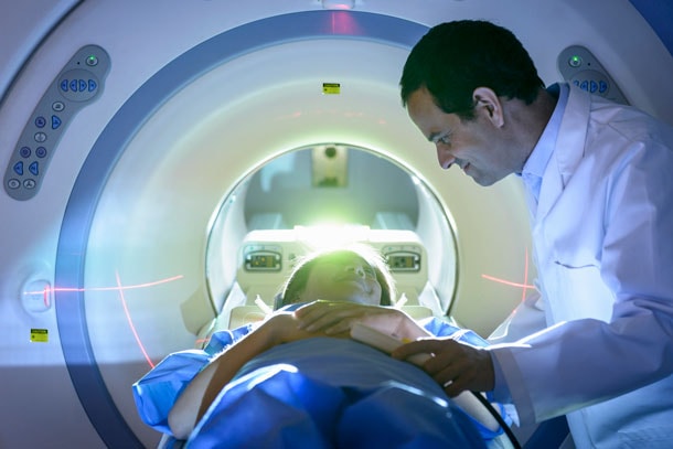
Magnetic resonance imaging uses radio waves and a strong magnetic field to create clear, detailed images of internal organs and tissues. Because X-rays are not used, no radiation exposure is involved. Instead, radio waves are directed at the body's protons within the magnetic field. This exam takes 30 to 50 minutes on average and consists of several imaging series. Some studies will require a small intravenous injection of a contrast agent that usually contains the metal gadolinium. MRI contrast does not contain iodine, an element used in other contrast agents for X-rays or CT scans, so it may be safer for patients who are sensitive to iodine.
Magnetic Resonance Angiography (MRA)
MRA is a noninvasive procedure that uses MRI to visualize blood vessels as two-and-three-dimensional images that can be viewed on a computer monitor. This requires no X-rays, invasive catheter placement or iodinated contrast material, but does involve an intravenous injection of Gadolinium. MRA is a painless, shorter exam than a catheter angiography. The results can help determine whether surgery or treatment such as angioplasty is needed, and to plan that treatment.
Ultrasound (Sonography)
High frequency sound waves are used to see inside the body. A transducer — a device that acts like a microphone and speaker—is placed in contact with the body using a special gel that helps transmit the sound. As the sound waves pass through the body, echoes are produced and bounce back to the transducer. By reading the echoes, the ultrasound can produce images that illustrate the location of a structure or abnormality, as well as provide information about its composition. Ultrasound is a painless way to examine the heart, liver, pancreas, spleen, blood vessels, breast, kidney or gallbladder and is a crucial tool for obstetrics.
Bone Densitometry
Both men and women can develop osteoporosis, although it's more common in women, particularly after menopause. Talk to your doctor about risk factors of osteoporosis. Diagnosed early, it can be both treatable and preventable. NNMC offers low-dose X-ray bone density screening, which is one of the best methods for early detection.
Mammography
The most widely used and recognized imaging method for the detection of breast cancer is a mammogram. This low radiation X-ray often can detect abnormalities in the breast before anything can be felt. Though digital mammography is a very effective method for detecting breast cancer, in certain cases, additional imaging tests are needed for complete evaluation. Therefore, additional tests such as ultrasound, MRI or other diagnostic imaging may be recommended. Women over 40—or younger women with a family history of breast cancer — should have annual mammograms.
Angiography
Angiography is an imaging procedure that takes images of blood vessels in various parts of the body, such as the brain, neck, abdomen, legs, kidneys and heart. This imaging helps doctors to determine whether vessels are diseased, narrowed, enlarged or blocked. There are three major forms of angiography: catheter angiography, computed tomography angiography (CTA) and magnetic resonance angiography (MRA).
Catheter Angiography
A catheter is usually inserted into an artery in the groin and advanced into the area of the circulatory system being examined. Imaging is performed using X-rays, and contrast material is often sent through the catheter to highlight the vessels while the X-rays are being taken. This process is used widely as a preoperative procedure and as a guide to perform angioplasty or stent placement.
Cardiac Catheterization and Percutaneous Coronary Intervention (PCI)
This technology provides faster and more precise cardiovascular exams, and can potentially save the heart muscle from damage or death. Coronary angioplasty or stent placement can allow blood to return to areas threatened by narrowed or blocked arteries.
Electrophysiology Studies (EPS)
Electrophysiology studies test the electrical activity of your heart to find where an abnormal heartbeat is coming from. Heart attacks, aging and high blood pressure may cause scarring of the heart leading to an irregular heartbeat pattern. In addition, EPS can be used by physicians to see the impact medicine is having in the treatment of arrhythmia, to determine if a pacemaker might help you and if you are at risk for additional heart problems such as fainting and cardiac arrest. The physician inserts one or more EP catheters into veins in your groin, arm or neck and threads them into the heart. Electrodes at the tip of catheters collect information about your heart's electrical activity and treat areas of your heart that are causing the problem.
Prestigious ACR and TJC Accreditation
Northern Nevada Medical Center has achieved the high standards required for American College of Radiology (ACR) accreditation in all imaging areas, including nuclear medicine, mammography, CT, MRI and both diagnostic and vascular ultrasound. ACR accreditation is a voluntary process in which a facility allows all aspects of its imaging procedures to be rigorously assessed by independent analysts.
These analysts evaluate image quality, equipment performance, policies, procedures and personnel qualifications. The ACR grants accreditation to facilities for establishing and maintaining high practice standards. These standards were designed to enhance the practice of medical imaging and the delivery of comprehensive healthcare services by board-certified physicians and medical physicists who are experts in their fields.
Northern Nevada Medical Center is also accredited by The Joint Commission (TJC), the gold seal of approval in healthcare certification, signifying adherence to the highest levels of technical quality, patient care and safety.
Contact Information
For Diagnostic Imaging scheduling, call 775-356-4042.
Diagnostic Imaging at Northern Nevada Medical Center
2375 East Prater Way
Sparks, NV 89434
Monday - Saturday, 7:30 a.m. – 5 p.m.

